سلام دکتر عزیزم خسته نباشید
این سونو خواهرمه.
4 روز قبل از پریودش داده.
دکترش دو ماه دیگه بهش نوبت داده.
پرسش (1404/08/13):
سلام دکتر جان این سونو رو میشه تفسیر کنید.
 182
182 10
10 1404/08/13
 الی ام
 0
0 1404/08/13
 الی ام
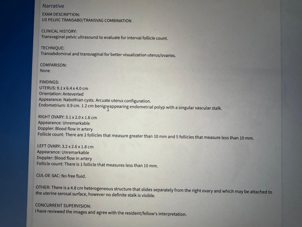
IMPRESSION: There is a 4.8 cm right paraovarian structure without a definite stalk connecting it to the uterus. Differential considerations include a pedunculated uterine fibroid with a nonvisualized stalk versus a round ligament fibroid. Malignancy is felt to be less likely, but given patient history and expressed concerns, MRI of the pelvis can be considered for further evaluation. This finding and these recommendations were discussed with the patient by Dr. Rockwell at the time of exam. Arcuate uterus configuration. Follicle count: - Right ovary: 2 follicles measuring greater than 10 mm, 5 follicles measuring less than 10 mm - Left ovary: 1 follicle measuring less than 10 mm
Narrative
EXAM DESCRIPTION: US PELVIC TRANSABD/TRANSVAG COMBINATION CLINICAL HISTORY: Transvaginal pelvic ultrasound to evaluate for interval follicle count. TECHNIQUE: Transabdominal and transvaginal for better visualization uterus/ovaries. COMPARISON: None FINDINGS: UTERUS: 9.1 x 6.4 x 4.0 cm Orientation: Anteverted Appearance: Nabothian cysts. Arcuate uterus configuration. Endometrium: 0.9 cm. 1.2 cm benign-appearing endometrial polyp with a singular vascular stalk. RIGHT OVARY: 3.1 x 2.0 x 1.6 cm Appearance: Unremarkable Doppler: Blood flow in artery Follicle count: There are 2 follicles that measure greater than 10 mm and 5 follicles that measure less than 10 mm. LEFT OVARY: 3.2 x 2.6 x 1.8 cm Appearance: Unremarkable Doppler: Blood flow in artery Follicle count: There is 1 follicle that measures less than 10 mm. CUL-DE-SAC: No free fluid. OTHER: There is a 4.8 cm heterogeneous structure that slides separately from the right ovary and which may be attached to the uterine serosal surface, however no definite stalk is visible. CONCURRENT SUPERVISION: I have reviewed the images and agree with the resident/fellow's interpretation.
 0
0 1404/08/13
 الی ام
گویا خواهرم در مورد این فیبروم خیلی اظهار نگرانی کرده و دکترم به همین خاطر توصیه به ام آر ای کرده.
ممنون میشم نظرتون رو بدونم💗🤍😘
ممنون میشم نظرتون رو بدونم💗🤍😘
 0
0 1404/08/13
 پزشک اوما
سلام به روی ماهتون عزیزم
لطفا عکس گزارش را ارسال کنید تا ببینم
لطفا عکس گزارش را ارسال کنید تا ببینم
 0
0 1404/08/13
 الی ام

 0
0 1404/08/13
 الی ام
دکتر جان این به درد میخوره؟
عاخه ایران نیس ریپورت کاغذی ندادن
عاخه ایران نیس ریپورت کاغذی ندادن
 0
0 1404/08/15
 پزشک اوما
بله جانم خوب است
کیست های نابوتین دیده شده اهمیت ندارد و اقدام لازم ندارد
رحم ارکوییت یا خمیده هست، که ممکن است گاها خطر زایمان زودرس یا سقط مکرر را بالا ببرد ولی ابن تشخیص قطعیت ندارد و لزوما منتهی به این مشکلات هم نمیشود
در کل شواهدی به نفع بدخیمی بنظر وجود ندارد جانم و بله بنظر فیبروم پایه دار دارند ولی قطعا انجام mri تصویر دقیق تری میدهد بنظز من اگر همکاری تیم پزشکیررا دارند حتما انجام دهند
 0
0 1404/08/15
 پزشک اوما
ولی مجموعا شواهد نگران کننده من نمیبینم
 0
0 1404/08/15
 الی ام
ممنونم ازتون دکتر جان🌹😘
شاد و برقرار باشید
شاد و برقرار باشید
 0
0 1404/08/15
 پزشک اوما
خواهش میکنم گل من ❤️😘❤️ار دعای خیلی زیباتون ممنونم و الهی که همیشه شاد باشید 😘💕💜
 0
0 1404/08/15
 پزشک اوما
stk: |~|11|~|103_col
سوالات مشابه
- سونوگرافی 35 هفته بارداری
- تفسیر سونو غربالگری اول
- تفسیر سونو و ازمایش غربالگری اول
- زمان زایمان طبق پریود یا آن تی
- سلام آیا سونو مشکل داره ؟؟؟
- سونو واژینال طول سرویکس در سی هفته
- شرایط سونو واژینال چجوریه
- کسی بیداره عایاااا؟ میخوام برم ازمایش هورمونی بدم بیاین کمک سوال دارم😂😂😂
- سونو تشکیل قلب
- لطفا سونوی منو تفسیر کنید خانم دکتر عزیزم


